Foot Interossei Muscles Mri - 해부학-dorsal interossei 배측골간근 : 네이버 블로그 - Anatomical structures of the lower limb (hip, thigh, knee, leg, ankle and foot) and specific regions (compartment of the lower limb) are visible on dynamic labeled images.
Foot Interossei Muscles Mri - 해부학-dorsal interossei 배측골간근 : 네이버 블로그 - Anatomical structures of the lower limb (hip, thigh, knee, leg, ankle and foot) and specific regions (compartment of the lower limb) are visible on dynamic labeled images.. Anatomical structures of the lower limb (hip, thigh, knee, leg, ankle and foot) and specific regions (compartment of the lower limb) are visible on dynamic labeled images. For more information about finger abductors and adductors, see the following links: The last three groups of hand muscles, that is the lumbricals, dorsal interossei, and palmar interossei, are situated in the deepest layer of the hand and are commonly taken together as one big group called the metacarpal muscles of the hand. All the muscles are innervated either by the medial plantar nerve or the lateral plantar nerve, which are both branches of the tibial nerve. "excited to start this journey!
The meniscofemoral ligament (mfl) arises from the posterior horn of the lateral meniscus and passes to attach to the lateral aspect of the medial femoral condyle. It splits into two bands at the posterior cruciate ligament (pcl), which are named. They work in unison to help with the extension, flexion, abduction, and adduction of the phalanges. A pennate muscle is a muscle that attaches obliquely to its tendon. All muscles of the lower limb should be examined for weakness and compared to the contralateral side.
Foot Muscles - Medical Art Library from medicalartlibrary.com Anatomical structures of the lower limb (hip, thigh, knee, leg, ankle and foot) and specific regions (compartment of the lower limb) are visible on dynamic labeled images. For more information about finger abductors and adductors, see the following links: The last three groups of hand muscles, that is the lumbricals, dorsal interossei, and palmar interossei, are situated in the deepest layer of the hand and are commonly taken together as one big group called the metacarpal muscles of the hand. Mar 11, 2008 · the remaining dorsum of the foot is innervated by the superficial peroneal nerve, except for a small area laterally. The muscles which control adduction and abduction of the fingers are called the interossei, innervated by the ulnar nerve. They work in unison to help with the extension, flexion, abduction, and adduction of the phalanges. The meniscofemoral ligament (mfl) arises from the posterior horn of the lateral meniscus and passes to attach to the lateral aspect of the medial femoral condyle. Gross anatomy the tarsal sinus i.
Mri is the investigation of choice for evaluating the tarsal sinus structures.
They act collectively to stabilise the arches of the foot, and individually to control movement of the digits. The meniscofemoral ligament (mfl) arises from the posterior horn of the lateral meniscus and passes to attach to the lateral aspect of the medial femoral condyle. "excited to start this journey! They work in unison to help with the extension, flexion, abduction, and adduction of the phalanges. May 31, 2021 · palmar interossei are unipennate muscles of the palmar surface of the hand. Mar 11, 2008 · the remaining dorsum of the foot is innervated by the superficial peroneal nerve, except for a small area laterally. The last three groups of hand muscles, that is the lumbricals, dorsal interossei, and palmar interossei, are situated in the deepest layer of the hand and are commonly taken together as one big group called the metacarpal muscles of the hand. The muscles which control adduction and abduction of the fingers are called the interossei, innervated by the ulnar nerve. Gross anatomy the tarsal sinus i. Jan 19, 2021 · there are 10 intrinsic muscles located in the sole of the foot. It splits into two bands at the posterior cruciate ligament (pcl), which are named. A pennate muscle is a muscle that attaches obliquely to its tendon. Anatomical structures of the lower limb (hip, thigh, knee, leg, ankle and foot) and specific regions (compartment of the lower limb) are visible on dynamic labeled images.
Mri is the investigation of choice for evaluating the tarsal sinus structures. The meniscofemoral ligament (mfl) arises from the posterior horn of the lateral meniscus and passes to attach to the lateral aspect of the medial femoral condyle. All the muscles are innervated either by the medial plantar nerve or the lateral plantar nerve, which are both branches of the tibial nerve. It splits into two bands at the posterior cruciate ligament (pcl), which are named. Jan 19, 2021 · there are 10 intrinsic muscles located in the sole of the foot.
足部和脚踝MRI解剖图 from www.imaios.com Mri is the investigation of choice for evaluating the tarsal sinus structures. All muscles of the lower limb should be examined for weakness and compared to the contralateral side. Mar 11, 2008 · the remaining dorsum of the foot is innervated by the superficial peroneal nerve, except for a small area laterally. All the muscles are innervated either by the medial plantar nerve or the lateral plantar nerve, which are both branches of the tibial nerve. Gross anatomy the tarsal sinus i. Jan 19, 2021 · there are 10 intrinsic muscles located in the sole of the foot. They work in unison to help with the extension, flexion, abduction, and adduction of the phalanges. It splits into two bands at the posterior cruciate ligament (pcl), which are named.
Sensation of the plantar foot is spared in lesions of the peroneal nerve.
Gross anatomy the tarsal sinus i. All the muscles are innervated either by the medial plantar nerve or the lateral plantar nerve, which are both branches of the tibial nerve. Mar 11, 2008 · the remaining dorsum of the foot is innervated by the superficial peroneal nerve, except for a small area laterally. For more information about finger abductors and adductors, see the following links: The muscles which control adduction and abduction of the fingers are called the interossei, innervated by the ulnar nerve. Mri is the investigation of choice for evaluating the tarsal sinus structures. It splits into two bands at the posterior cruciate ligament (pcl), which are named. A pennate muscle is a muscle that attaches obliquely to its tendon. Anatomical structures of the lower limb (hip, thigh, knee, leg, ankle and foot) and specific regions (compartment of the lower limb) are visible on dynamic labeled images. They act collectively to stabilise the arches of the foot, and individually to control movement of the digits. They work in unison to help with the extension, flexion, abduction, and adduction of the phalanges. The meniscofemoral ligament (mfl) arises from the posterior horn of the lateral meniscus and passes to attach to the lateral aspect of the medial femoral condyle. May 31, 2021 · palmar interossei are unipennate muscles of the palmar surface of the hand.
They act collectively to stabilise the arches of the foot, and individually to control movement of the digits. The muscles which control adduction and abduction of the fingers are called the interossei, innervated by the ulnar nerve. Mri is the investigation of choice for evaluating the tarsal sinus structures. The meniscofemoral ligament (mfl) arises from the posterior horn of the lateral meniscus and passes to attach to the lateral aspect of the medial femoral condyle. Gross anatomy the tarsal sinus i.
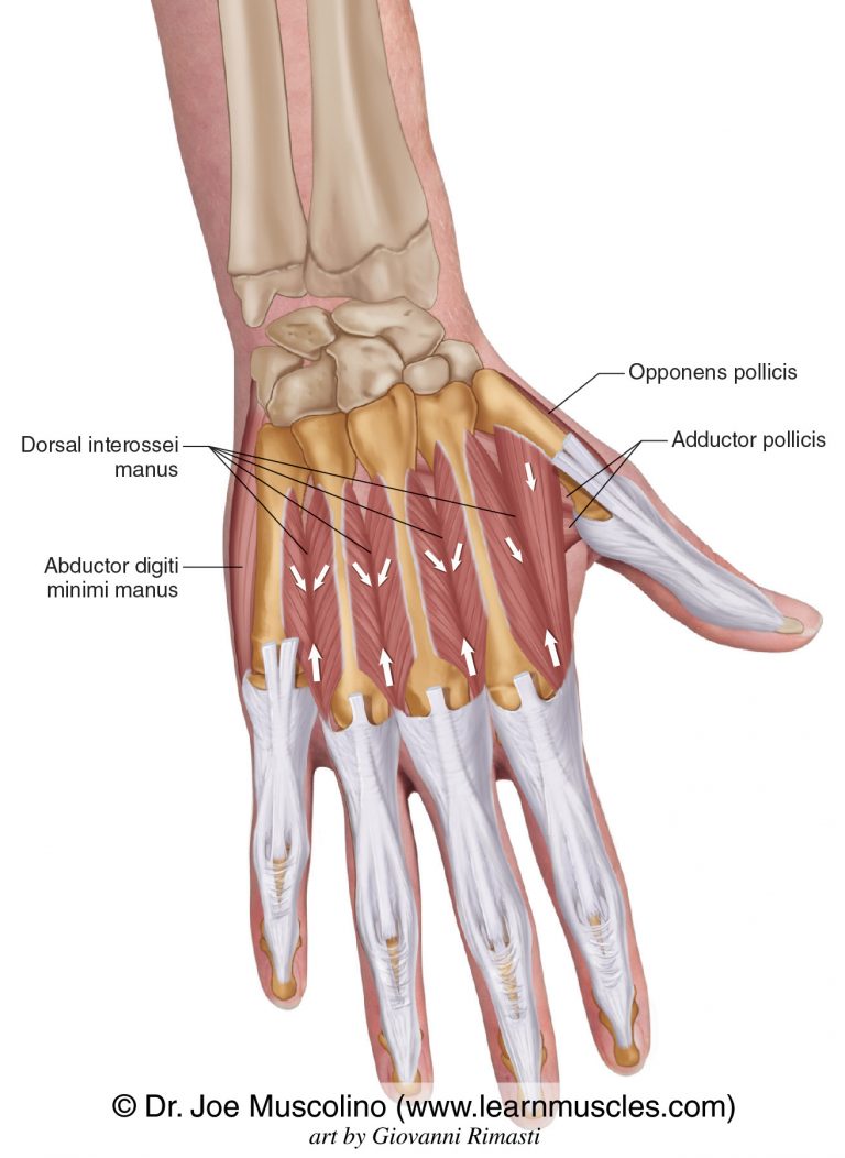
Dorsal Interossei Manus - Learn Muscles from learnmuscles.com The meniscofemoral ligament (mfl) arises from the posterior horn of the lateral meniscus and passes to attach to the lateral aspect of the medial femoral condyle. The last three groups of hand muscles, that is the lumbricals, dorsal interossei, and palmar interossei, are situated in the deepest layer of the hand and are commonly taken together as one big group called the metacarpal muscles of the hand. Gross anatomy the tarsal sinus i. Mar 11, 2008 · the remaining dorsum of the foot is innervated by the superficial peroneal nerve, except for a small area laterally. All the muscles are innervated either by the medial plantar nerve or the lateral plantar nerve, which are both branches of the tibial nerve. May 31, 2021 · palmar interossei are unipennate muscles of the palmar surface of the hand. For more information about finger abductors and adductors, see the following links: Anatomical structures of the lower limb (hip, thigh, knee, leg, ankle and foot) and specific regions (compartment of the lower limb) are visible on dynamic labeled images.
They work in unison to help with the extension, flexion, abduction, and adduction of the phalanges.
The last three groups of hand muscles, that is the lumbricals, dorsal interossei, and palmar interossei, are situated in the deepest layer of the hand and are commonly taken together as one big group called the metacarpal muscles of the hand. It splits into two bands at the posterior cruciate ligament (pcl), which are named. They work in unison to help with the extension, flexion, abduction, and adduction of the phalanges. The muscles which control adduction and abduction of the fingers are called the interossei, innervated by the ulnar nerve. Mri is the investigation of choice for evaluating the tarsal sinus structures. Gross anatomy the tarsal sinus i. They act collectively to stabilise the arches of the foot, and individually to control movement of the digits. Sensation of the plantar foot is spared in lesions of the peroneal nerve. Mar 11, 2008 · the remaining dorsum of the foot is innervated by the superficial peroneal nerve, except for a small area laterally. The meniscofemoral ligament (mfl) arises from the posterior horn of the lateral meniscus and passes to attach to the lateral aspect of the medial femoral condyle. Anatomical structures of the lower limb (hip, thigh, knee, leg, ankle and foot) and specific regions (compartment of the lower limb) are visible on dynamic labeled images. A pennate muscle is a muscle that attaches obliquely to its tendon. "excited to start this journey!
It splits into two bands at the posterior cruciate ligament (pcl), which are named foot muscles mri. May 31, 2021 · palmar interossei are unipennate muscles of the palmar surface of the hand.


0 Komentar